


Five to ten percent of blindness globally is caused by the uncommon inflammatory eye disease uveitis. The need for immunosuppressive treatment and severe disease progression are commonly linked to posterior uveitis in particular. The underlying choroid, which supplies the retina with nutrition, as well as the retina itself become inflamed in posterior uveitis. Researchers at the University of Bonn’s Department of Ophthalmology looked at color-coded fundus autofluorescence as a potential novel diagnostic tool. The kind of uveitis may be identified by the retina’s fluorescence. This is a crucial prerequisite for accurate illness diagnosis and treatment.
Uveitis, a Common Eye Condition
Scientific Reports reported the results of the study. The uncommon condition posterior uveitis causes blurred vision, floaters, and odd light perception, but no discomfort in people who have it. “However, there might be dire repercussions: Uveitis accounts for five to ten percent of blindness globally. Uveitis is an uncommon condition, but posterior uveitis in particular may have a bad prognosis and frequently needs immunosuppressive treatment, according to Dr. Maximilian Wintergerst of the University of Bonn’s Ophthalmology Department.
The sickness manifests itself in many ways. The retina or choroid in the eye becomes inflamed in posterior uveitis. The choroid provides nutrition to the retina’s outer layers while the retina transforms the incoming light into nerve impulses.
The many subtypes of uveitis are difficult to identify from one another, according to Wintergerst. However, since various subtypes frequently require various therapy modalities, a precise diagnosis is even more crucial. To help with the diagnosis of posterior uveitis, researchers from the University of Bonn’s Department of Ophthalmology, along with peers from the University Hospital Bonn’s Medical Biometry and Rheumatology Departments and the University Hospital of Ophthalmology in Bern (Switzerland), have looked into a new imaging technique.
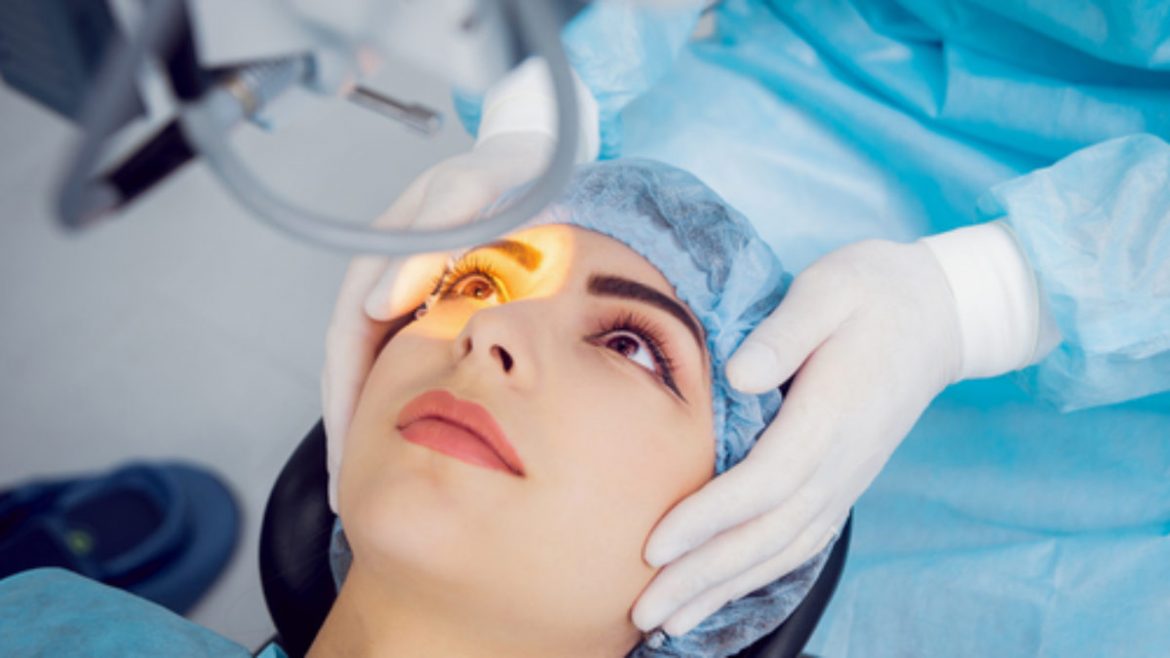
Color-coded fundus autofluorescence was assessed by the team (Spectrally Resolved Autofluorescence Imaging). The newly created apparatus for the tests was given to the researchers by the CenterVue (iCare) firm of Padua (Italy). The retina is illuminated during this procedure using blue light.
The retina reflects light at a different wavelength after it has been absorbed by it. The instrument divides the signals into a green and a red component before measuring the fluorescence.
According to Wintergerst, “the particular posterior uveitis subtype implicated determines, among other parameters, the green-to-red ratio of the light emitted from each inflammatory centre.” 45 study participants had their eyes tested by the researchers. Prior to treatment, the precise subtype of uveitis was identified in each case. This comprised the results of laboratory tests, radiologic and serologic examinations, and occasionally genetic and multidisciplinary clinical evaluations.
Greater diagnostic accuracy using color-coded fundus autofluorescence
About 800 inflammatory foci in the patients’ eyes were examined by the researchers using the green-red ratio in the fundus fluorescence. According to Prof. Dr. Dr. Robert Finger, co-author of the study and director of the uveitis clinic at the University Hospital Bonn’s Department of Ophthalmology, “our results show that this ratio can be very characteristic and useful as a marker for differentiating the various posterior uveitis subtypes.”
It could enable us to make diagnoses with greater assurance in the future. This is a significant step for the University of Bonn’s Ophthalmology Department, especially as Finger collaborates with colleagues to oversee the country’s national uveitis registry, TOFU (Treatment exit options for non-infectious uveitis, www.tofu-uveitis-register.de).
Also Read: Children with Down syndrome like crunchy, fatty meals and detest sticky ones: Study
Long-term illness progression documentation and therapy suggestion development are the objectives.
According to department director Prof. Dr. Frank Holz, “In the current work, we offer the precise technological foundation of color-coded fundus autofluorescence in ophthalmology in partnership with our international colleagues.” In addition to more accurate diagnosis, this technique may in the future provide improved monitoring of posterior uveitis.
Follow Medically Speaking on Twitter Instagram Facebook