


A recent study from the University of South California demonstrates that a specific pattern of white matter connectivity has been found in the brains of autistic people. It is also distinct from the pattern seen in those with Developmental Coordination Disorder (DCD).
According to the study, which was published in the journal ‘Scientific Reports,’ roughly 85 percent of autistic persons have had or are at risk of being diagnosed with DCD, a disorder that interferes with learning and motor control. DCD can impede daily tasks such as typing, dressing, and walking, reducing one’s social involvement and enjoyment.
Distinguishing between the brain activity patterns of autism spectrum disorder (ASD) and DCD populations is critical because the widespread comorbidity of ASD and DCD confounds previous autism research, which was thought to be exclusively investigating its core social-communication symptoms at the time it was conducted.
“As the scientific community has learned more about DCD, we realised that white matter disparities previously observed in the autism literature might really be attributable to this underlying motor comorbidity,” stated senior author Lisa Aziz-Zadeh. “In reality, that’s exactly what our team discovered: many earlier research findings are likely not representing autism’s core symptoms, but rather co-occurring DCD.”
Aziz-Zadeh is an associate professor in the USC Dornsife College of Letters, Arts, and Sciences’ Brain and Creativity Institute and the Department of Psychology. She is the director of the USC Center for the Neuroscience of Embodied Cognition, which manages research projects funded by the National Institutes of Health, the US Department of Defense, and the Intelligence Advanced Research Projects Activity of the Office of the Director of National Intelligence.
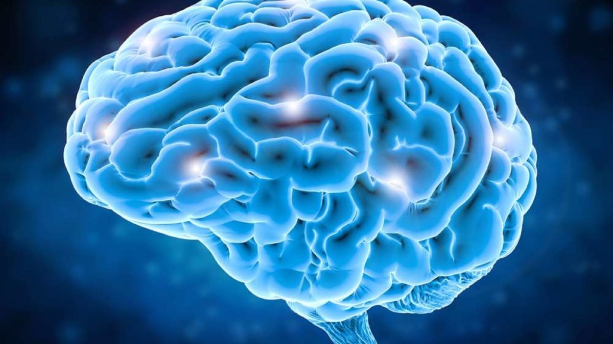
Aziz-Zadeh and colleagues employed diffusion weighted MRI, a method for evaluating functional brain connections, on children and teenagers aged 8 to 17 years old who were randomly allocated to one of three research groups: those with ASD, those with suspected DCD, and those who were normally developing.
The photos were evaluated, compared, and connected with physical and social behaviour assessments performed by the participants. The researchers discovered that numerous structural brain connection patterns previously associated with autism also overlap with DCD. The team was able to identify three white matter pathways with distinctly different connectivity, distinct from the DCD and typically developing groups: the longitudinal fibres and u-fibers of the mid-cingulum, the corpus callosum forceps minor/anterior commissure, and the left middle cerebellar peduncle. These disparities were also connected to autistic participants’ emotional performance and/or autism severity. Children with DCD had distinct white matter patterns in the left cortico-spinal and cortico-pontine tracts.
“These findings show that we can use advanced imaging to differentiate between autism’s hallmark social symptoms and other motor-related symptoms at the level of brain anatomy,” said Emily Kilroy, the study’s first author and a former postdoctoral scholar in Aziz-lab Zadeh’s during the data collection period. “Of course, people are much more than their brain structure,” says the researcher, “but this level of clarity and precision at the anatomical level brings us one step closer to understanding the molecular foundation and presentation of autism.”