


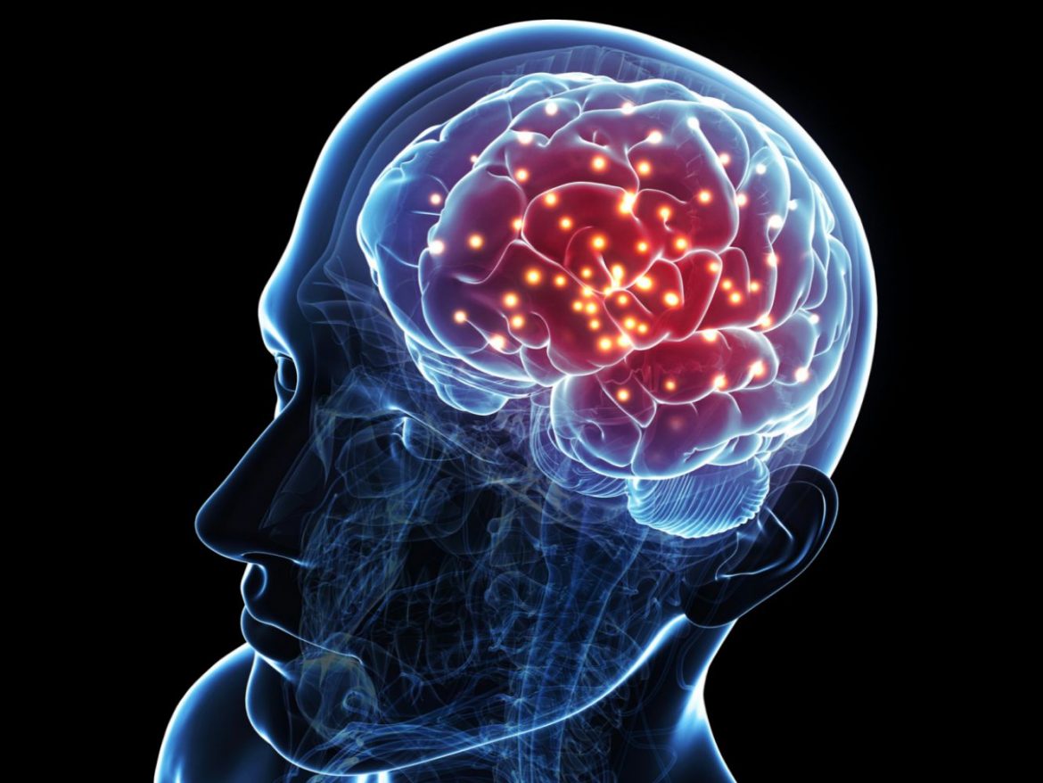
How do brain regions communicate is a perennial topic in neuroscience. The cause for this has now been identified by a new study at the Sainsbury Wellcome Centre.
The Sainsbury Wellcome Centre at UCL researchers employed causal approaches to discover how two neocortical parts of the brain connect with one another and discovered that their impact on each other varies over considerably quicker timeframes than previously imagined. Neuroscientists have struggled to disentangle the networks that give birth to behaviour in the brain, which has around 80 billion neurons and 100 trillion connections.
SWC researchers explain how two visual regions in the cerebral cortex, in a new study published today in Neuron.
SWC researchers reveal how two visual regions in the cerebral cortex, the primary visual cortex (V1) and lateromedial area (LM), impact one another and how this connection varies over time in a new study published today in Neuron.
“We sought to investigate inter-area communication to learn how various brain regions collaborate to comprehend visual inputs.” We know from previous research that there is a visual area hierarchy with feedforward and feedback pathways. “In primates, the first level of hierarchy in the cerebral cortex is V1, and the second level is V2, which is analogous to LM in mice,” explained Mitra Javadzadeh, Research Fellow at SWC and co-author of the work.
“Our expectation from the anatomical connections between V1 and LM is that the effect of neuronal activity in one area on another would be relatively constant; however, we were surprised to find it is dynamic and changes over time. These changes can happen very rapidly, within tens of milliseconds,” said Sonja Hofer, Group Leader at SWC and co-author of the paper.
Historically, scientists have recorded from different brain areas and used statistical correlations to infer how one area influences another. In this study, Javadzadeh and Hofer instead took a causal approach by using neuronal perturbations to study the dynamics of inter-areal interactions over time.
The neuroscientists recorded populations of neurons in V1 and LM in mice and used optogenetics to briefly silence the activity of one area and quantify how the activity increased or decreased in the other area. This showed them the contribution of the first area in shaping the firing rates of the second area.
Also Read: Glucose metres may soon be used to detect SARS-CoV-2 antibody levels
Javadzadeh and Hofer measured these contributions over time while these brain areas were processing visual information. Surprisingly, they found that the effect of manipulating one area on the activity in another varied overtime on a fast timescale. For example, a neuron in area V1 could decrease its activity in response to area LM at the one-time point but not be influenced by LM activity 100 milliseconds later.
Furthermore, if the visual input was behaviorally meaningful for the animal, such as predicting the presence of a reward, these changes in impact happened much faster.
The authors hypothesise that these quickly shifting impacts may allow cortical areas to govern distinct elements of processing in the downstream brain regions they influence over extremely short time spans. That is, the function that distinct regions play in moulding each other’s activity might be fluid and suited to the dynamic needs of behaviour.
In addition to investigating the function of these dynamic interactions, Javadzadeh and Hofer are collaborating with scientists at the Gatsby Computational Neuroscience Unit, which is housed in the same building as SWC, to better understand the processes that cause them.
Follow Medically Speaking on Instagram