


New Delhi – A multi-disciplinary team of doctors has treated an 18-month infant who was suffering from Trigonocephaly. A rare condition in which the shape of the front of the head is triangular, causing aesthetic and neuropsychological development defects. In such cases, the surgeries are challenging and it also requires pre-planning. This surgery is a cranio-facial surgery that was performed to correct the deformity by a team of doctors which is led by Dr. Sonal Gupta, Director, Neuro and Spine Surgery and Dr. Richie Gupta, Additional Director, Plastic, Aesthetic and Reconstructive Surgery from Fortis Shalimar Hospital
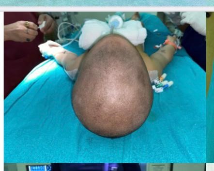
The infant when came for the treatment was having a triangular-shaped appearance on the forehead and hypotelorism,“The infant presented with a triangular-shaped appearance on the forehead and hypotelorism (a condition in which, the eyes appear closer together than normal). Normally, the bones forming the skull are separated by joints called sutures, which also serve as centers of growth. These sutures fuse at set times after birth, one of the earliest being the suture between frontal bones, the metopic suture which obliterates between 3-9 months of age. An early sutural fusion results in a lack of skull growth in a direction perpendicular to the suture. However, the brain is still growing at a rapid rate during this period and needs space to do so. Therefore, the condition needed to be corrected, as it could hamper the neuro-psychological development of the child in addition to the obvious aesthetic defect.” said Dr Sonal Gupta
“It was a complex surgery which took seven hours. To put an infant under anaesthesia for that long is a challenge. It was an intensive and precise process which would not have been possible without the support of the anaesthesiology team led by Dr Umesh Deshmukh (Director and HOD anaesthesiology). There was a risk of significant blood loss during the surgery, so multiple blood products were arranged. After the surgery, we shifted the patient to the ICU, where he stayed for 3 days under the care of Dr Amit Singh (Consultant Paediatric Critical Care). Post this period, he was shifted to the ward for 2 days and on the 6th day he was discharged. Even though I had first met with this patient when he was only two months old, I chose not to the surgery immediately as it would not have yielded the desired results.” Dr Sonal Gupta adds
According to Dr Richie Gupta the cranio-facial surgery is needed to correct the deformity, “A cranio- facial approach was needed to correct the deformity. We fed the digital data from the patient’s 3DCT scan into the 3D printer to print two rapid prototype skull and upper face models, which were exact replicas of the patient’s skull. We worked on these to determine the exact site, dimensions, and angles of the osteotomies (bony cuts) on the patient’s skull. There was no scope for even the smallest of deviations as it could cause the child to go blind, bleed profusely, or even suffer from brain damage. To ensure that the actual surgery went off smoothly, we conducted the mock surgery on the 3D model of the skull a day prior.”