


Nobody enjoys having biopsies or having endless tests performed on them, even if they are necessary for keeping track of their health. According to Japanese experts, a revolutionary technology that might greatly minimise the invasiveness of cancer diagnostics has just been discovered.
In a study published in the Journal of the American Chemical Society in September, researchers from Tokyo Medical and Dental University (TMDU) revealed a novel approach for identifying the cancer-related marker using breast cancer cell lines.
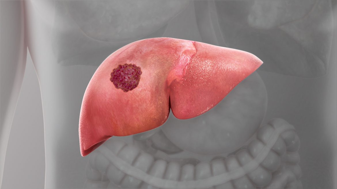
Finding cancer-related indicators is a highly effective method for determining diagnosis, prognosis, and response to treatment. These markers may now be found in patient samples like blood and urine thanks to modern technology, offering a non-invasive way to monitor and assess patients.
“Circulating tumour cells (CTCs), which are cancer cells found in the blood, are one of the main targets used to evaluate cancer patients’ blood samples,” states Miyuki Tabata, first author of the study. “However, it can be challenging to isolate these cells from the blood, and current approaches do not adequately detect both epithelial cell and mesenchymal cell markers, which are important for determining the stage of cancer.”
The researchers employed a device known as an ion-sensitive field effect transistor (ISFET), which is a small electrical circuit that is activated by a change in pH, to develop a system that can swiftly and readily identify cancer-related markers on CTCs (and maybe other elements in the blood). These transistors were coated with breast cancer cells, and an antibody connected to a chemical reporter that changes pH when the antibody binds to the cells was then added.
“We found that the chemical reporter glucose oxidase successfully detected the expression of epidermal growth factor receptor (EGFR), a marker of poor cancer prognosis, on CTC membranes,” says Yuji Miyahara, senior author of the study. “Furthermore, the strength of the chemical signal correlated with the amount of EGFR expressed by the cells.”
Importantly, these outcomes lined up with the EGFR levels found using other methods, showing that the ISFET strategy correctly detects the expression of cancer-related markers on cells.
“These results provide proof of concept that an ISFET-based system can be used to assess patient cancer status based on liquid biopsy samples efficiently,” states Tabata.
Because of the capacity to create ISFETs the size of a single cell and combine them into massive arrays, this technique can enable high-throughput study of cancer cells at single-cell resolution. Furthermore, the employment of different antibodies for the chemical enzyme detection phase may allow for the simultaneous investigation of many cancer-related markers.
Also Read: New evidence firmly revives Covid Wuhan lab origin theory: Report