


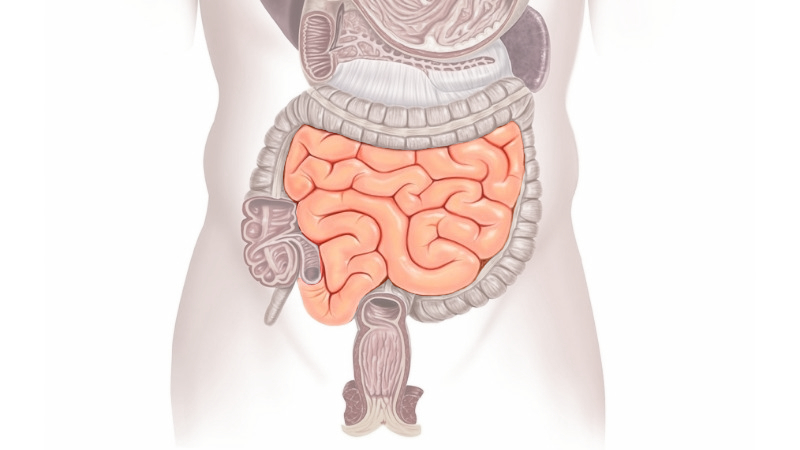
A component in the intestine that plays an important function in healing damaged tissue has been uncovered by researchers in a recent study.
The findings were published in the journal Cell Stem Cell by researchers from Cedars-Sinai and the University of California, San Francisco (UCSF). Endothelial cells in lymphatic veins create chemicals that are required for the maintenance and control of stem cells and tissues in the colon, according to researchers. These lymphatic endothelial cells live in close proximity to specific stem cell niches, which are microenvironments that promote stem cell regeneration.
Cell Stem Cell, a peer-reviewed publication, reported the findings.
“It’s important for us to understand niches and how lymphatics communicate with stem cells as part of the niche,” ANI quoted Ophir Klein, MD, PhD, senior author of the study and executive director of Cedars-Sinai Guerin Children’s. “Deciphering the mechanisms that explain how the ecosystem that supports stem cells works will help to lay the foundation for future discoveries that could one day lead to therapeutic strategies to repair damaged tissue.”
The gut is constantly renewed to survive the wear and tear caused by food breakdown and the presence of waste, which can harm and kill cells. The gut must continually renew itself with healthy cells, and thankfully, it has an amazing potential for cell regeneration.
The division of intestinal stem cells to produce additional cells is governed by their surrounding niche, which is made up of several cell types and serves as a vital source of signals. However, it is unknown which niche cells provide signals throughout various stages of damage.
To get a better understanding of stem cell activity, the researchers aimed to find out which cells, namely lymphatic endothelial cells, aid in the repair of intestinal epithelial cells.
“Lymphatics are in very close proximity to the stem cells, and almost all the stem cell compartments are near lymphatics,” said Brisa Palikuqi, PhD, co-first author of the study and a postdoctoral fellow in the Klein Laboratory at UCSF. “Because the two cell types are in such proximity, this made us believe that these lymphatics may play an important role.”
Lymphatics express several factors, including a gene, Rspo3, that is known to be important for stem cells to function. To determine whether the gene played an important role in intestinal stem cell regulation, the investigators deleted the gene in mice and then used single-cell sequencing to see how stem cells in the intestine would react without Rspo3.
Also Read: India records two more monkeypox cases
Initially, the stem cells did not have any response to the change in the environment. The researchers then decided to injure the system by delivering a chemotherapy drug that kills any proliferating cells wherever the drug travels through the body.
“When we did this, all of a sudden the stem cells and the intestine had to proliferate a lot more and replace a lot more cells on a daily basis,” said Jeremie Rispal, PhD, also a postdoctoral researcher in the Klein Laboratory at UCSF and the other co-first author of the study.
The loss of the Rspo3 gene led to a lower number of stem and progenitor cells, hindering recovery after the injury.
“This discovery showed us that lymphatic endothelial cells are a key component of the intestinal niche and essential for intestinal repair after cases of damage, like chemotherapy,” said Klein, who also holds the David & Meredith Kaplan Distinguished Chair in Children’s Health at Cedars-Sinai.
Klein noted that the study demonstrates how lymphatic endothelial cells play a much larger role than previously thought in stem cell regeneration and may play a role in disease, perhaps even influencing the promotion of cancer.
“We are just beginning to understand the functions of the lymphatic vasculature,” Klein said.
Klein previously directed the Institute for Human Genetics and served as chief of the Divisions of Medical Genetics and Craniofacial Anomalies at UCSF, where he remains an adjunct professor. Klein conducted the study at both USCF and Cedars-Sinai.
Follow Medically Speaking on Twitter Instagram Facebook